• What is CT Scanning of the Head?
• What are some common uses of the procedure?
• How should I prepare for the CAT scan?
• How does the procedure work?
• Who interprets the results and how do I get them?
• What are the benefits vs. risks?
• What are the limitations of CT Scanning of the Head?
What is CT Scanning of the Head?
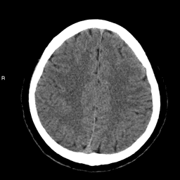
Computed tomography (CT), sometimes called CAT scan, uses special x-ray equipment to obtain many images from different angles, joining them together to show a cross-section of your brain. CT scanning provides detailed information on head injuries, stroke, brain tumors and other brain diseases. This imaging technique details bone, soft tissues and blood vessels. CT of the head/ brain is a patient-friendly exam that involves radiation exposure.
What are some common uses of the procedure?
- Detection of bleeding, brain damage and skull fractures in patients with head injuries.
- Detecting a blood clot or bleeding within the brain shortly after a patient exhibits symptoms of a stroke.
- Detection of stroke.
- Evaluation of the extent of bone and soft tissue damage in patients with facial trauma.
- Detection of bleeding in a patient with a sudden severe headache who may have a ruptured or leaking aneurysm.
- Detection of most brain tumors.
- Diagnosing diseases of the temporal bones, which may be causing hearing/balance problems or tinnitis.
- Detection of enlarged brain cavities (ventricles) in patients with hydrocephalus.
- Determining whether infection/inflammation or other changes are present in the paranasal sinuses.
- Planning radiation therapy for cancer.
- Non-invasive assessment of aneurysms or arteriovenous malformations through a technique called CT angiography.
- Detecting diseases or malformations of the skull.
- Three-dimensional imaging of the skull and brain structures for surgical planning.
How should I prepare for the CAT scan?
You should wear comfortable, loose-fitting clothing for your CT exam. Anything that might interfere with imaging of the head (especially metal)-such as earrings, eyeglasses, dentures, dental implants or hairpins-should be removed. No special preparation is needed for a CT scan of the head unless you are to receive a contrast material-a substance that highlights the brain and its blood vessels and makes abnormalities easier to see. CT scan contrast materials contain iodine, which can rarely cause a reaction in persons who are allergic. If the radiologist believes that an intravenous (IV) injection of a contrast material will be helpful, you may be asked in advance whether you have had an allergic reaction to contrast in the past. The radiologist should also know if you have asthma, multiple myeloma or any disorder of the heart or kidneys, or if you have diabetes – particularly if you are taking Glucophage (Metformin). You will be asked to sign an informed consent before having a CT with injection of a contrast material. You can see a copy of the consent on our website, under “forms” which will discuss the risks of the contrast. Women should always inform their doctor or x-ray technologist if there is any possibility that they are pregnant. In some cases an alternate study may be performed to reduce or eliminate the radiation exposure to the fetus.
Unlike conventional x-rays, which produce one dimensional images of the body, CT scanning uses x-rays in a much different way. In CT of the head, numerous x-ray beams are passed through the skull and brain at different angles, and special sensors measure the amount of radiation absorbed by different tissues (and lesions such as a bleeding or a tumor). As you lay still, the scanner parts revolve around you (although you cannot see this happen), emitting and recording x-ray beams from as many as a thousand points on the circle. A special computer program then uses the differences in x-ray absorption to form cross-sectional images, or "slices" of the brain. These slices are called tomograms, hence the name "computed tomography." The images can be reconstructed in any two-dimensional plane or into three-dimensional images.
Who interprets the results and how do I get them?
A neuroradiologist, who is a physician experienced in head, neck and spine CT examinations, will analyze the images and send a report with his or her interpretation to your personal physician. A preliminary interpretation may be available shortly after the exam with the complete report sent to the referring physician within twenty-four hours. Your primary physician or the radiologist may discuss the findings of the CT examination with you. New technology also allows for distribution of diagnostic reports and images over the Internet from our facility to your doctor.
What are the benefits vs. risks?
Benefits:
- CT of the head is now widely available and is performed in a relatively short time, at a reasonable cost-compared with MR imaging.
- CT scanning demonstrates changes in bone better than any other imaging method.
- CT imaging readily detects brain hemorrhage.
- The exam is usually used as an initial study for stroke detection.
- It provides detailed images of bone, soft tissue and blood vessels.
- CT is the method of choice for rapidly screening trauma victims to detect internal bleeding or other life-threatening conditions.
- CT Angiography depicts blood vessels supplying the brain, revealing aneurysms and vessel narrowing/occlusion.
Risks:
- CT involves exposure to radiation in the form of x-rays, but the benefit of an accurate diagnosis far outweighs the risk. The effective radiation dose from this procedure is about 2 mSv, which is about the same as the average person receives from background radiation in the general environment over a period of eight months.
- Women should always inform their doctor and x-ray technologist if there is any possibility that they are pregnant.
- Nursing mothers should wait 24 hours after contrast injection before resuming breast feeding.
- The risk of serious allergic reaction to iodine containing contrast material is rare. Personnel working at CT units are well equipped to deal with contrast reactions.
What are the limitations of CT Scanning of the Head?
Compared to MR imaging, the precise details of soft tissue (particularly the brain) are less visible on CT scans. CT is not sensitive in detecting inflammation of the meninges-the membranes covering the brain.
Compared to conventional angiography, computed tomography angiography (CTA) may, in some cases, not be as sensitive in the detection of aneurysms and arteriovenous malformations of the brain.
Copyright © 2007 Radiological Society of North America, Inc.
Source Info
© 2006 Mink Radiologic Imaging, Inc.